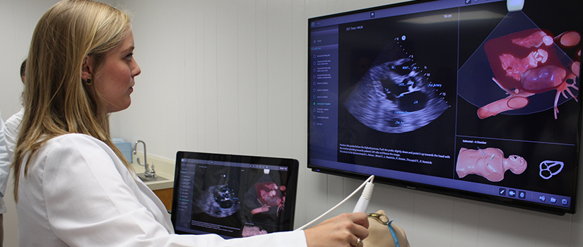
Skip to Content

School of Medicine Columbia

  • Student using ultrasound simulator

Integrated Ultrasound

This technology gives you the skills you need to make an impact in many settings, from the prominent technologically-advanced hospital to the rural primary care facility, and everywhere in between. 

In 2006, we became the first university in the country to integrate ultrasound technology throughout a four-year medical curriculum. Since then, we've worked with our partners to create curricula, test new technologies and encourage medical providers around the world to use ultrasound technology in ways that improve the quality of patient care. We believe ultrasound should be a core competency because there are practical applications in almost all medical disciplines. 

Back to the Bedside

The best place to teach the art, science and humanity of medicine is at the bedside. One of the benefits of ultrasound is that it provides you the opportunity to visit patients, to learn to talk to them and use the images as a teaching tool to help them understand their health. 

What You'll Learn

You’ll be exposed to ultrasound technology throughout the your curriculum.  The focus will be on the applications most relevant to specific courses and clerkships.

First Year Curriculum

 Small group introductory ultrasound sessions include: 

  • basic instrumentation and knobology.
  • image orientation.
  • hands-on scanning of neck vessels.

All education material is available to students online throughout all four years including: learning modules, videos, laboratory handouts and notes.

  1. Introduction to cardiac ultrasound (laboratory session)
    • Parasternal long axis view (PLAX) - B-mode only; identification of heart chambers, valves, review screen orientation and image orientation marker location, knobology, depth, focus, frequency, gain & adjustments
  2. Neck ultrasound ( laboratory session)
    • Carotid artery – B-mode and color flow mode – trace from common carotid to bifurcation, transverse  and longitudinal views, basic principles of color flow Doppler
    • Internal jugular vein – B-mode and color flow mode; anatomic differences of internal jugular vein  and carotid artery, shape, vessel wall, collapsibility, perform valsalva
    • Thyroid gland – B-mode; thyroid (both lobes and isthmus); echotexture, measurements, label structures, thyroid lobe volume estimation
  3. Kidney and bladder ultrasound (laboratory session)
    • Kidneys- Longitudinal and transverse views of right & left kidneys, obtain measurements
    • Urinary bladder – B-mode; identify bladder, measure bladder volume, note artifacts like posterior acoustic enhancement
    • Ureteric jets – Color flow mode; test of total ureteric obstruction
  4. Right upper quadrant (laboratory session)
    • Liver, gall bladder, right kidney, Morison’s pouch, diaphragm and right costophrenic angle
  5. Introduction to musculoskeletal ultrasound – the knee (laboratory session)
    • Anterior longitudinal suprapatellar view – patella, quadriceps tendon, femur, suprapatellar bursa
    • Suprapatellar transverse flexed knee view - quadriceps tendon, femoral condyles, articular cartilage
    • Infrapatellar longitudinal view – patellar ligament, fat pad, tibia
    • Anisotropy artifact   
  6. Brain and Cranial Nerves (presentation and demonstration) 
    • Ultrasound measurement of optic nerve sheath diameter for assessment of increased intracranial pressure
    • Ultrasound assessment of direct and consensual pupillary light reflex
    • Ultrasound assessment of ocular movement for patients with marked orbital swelling
    • Transcranial Doppler imaging of Circle of Willis vessels
  7. Ultrasound OSCE – proper transducer selection,  preset selection, probe orientation,  scan and identify selected anatomy from the five lab sessions; student is also evaluated on their interaction with the standardized patient
  1. Introduction to vascular ultrasound  (laboratory)
    • Vascular hemodynamics
    • Common carotid artery analysis
      • B-mode – transverse and longitudinal views
      • Color flow – direction of flow
      • Spectral Doppler/Pulse wave – measure velocity, Peak Systolic Velocity (PSV), End Diastolic Velocity (EDV), arterial and venous pulse wave forms
  2. Heart ultrasound – hemodynamics (laboratory)
    • Apical 4 chamber view (B-mode and color flow mode) – wall motion, valve motion, cardiac cycle with color flow
    • Heart sounds: Students work in pairs – one captures PLAX  view showing both the aortic valve and mitral valve while other student listens with stethoscope and notes relationship of heart sounds and valve closure. Students then reverse roles.
  3. Ultrasound in Shock (laboratory session)
    • Cardiac views for cardiogenic and obstructive shock
    • Specific views: PLAX,PSAX, apical 4-chamber, & subcostal 
    • Cardiomyopathy as a cause of cardiogenic shock – assess wall motion and shape of the left ventricle (LV) during cardiac cycle
    • Cardiac tamponade as a cause of obstructive shock – assess for pericardial effusion, the right ventricle (RV) size and compression with cardiac cycle
    • Pulmonary embolism as a cause of obstructive shock – assess for RV strain: size of RV and position of interventricular septum; assess RV and right atrium (RA) for thrombosis  
Ultrasound Assisted Physical Diagnosis :
  • Small group hands-on session
  • Students scan standardized patients to correlate physical exam findings with ultrasound findings
  • Diaphragmatic excursion, liver span, sonographic Murphy’s sign, heart sounds

Second Year Curriculum 

  1. Cardiac ultrasound   (laboratory session)
    • Standard cardiac views: Parasternal long and short axis views, apical 4 and 5 chamber, subcostal
    • Assess chambers, valves, wall thickness and motion
  2. Abdominal Aorta and Inferior VenaCava (IVC) assessment (laboratory session)
    • AAA screening; transverse and longitudinal, B mode, color flow, three measurements, characteristics that differentiate aorta from IVC
    • IVC – B mode and M Mode, measurement and IVC collapsibility index
  3. Lower extremity venous ultrasound (laboratory session)
    • Assess for deep venous thrombosis (DVT)  –  2 zone compression test of 1) the femoral vein at the saphenofemoral junction and 2) the popliteal vein, color flow Doppler, normal phasic venous flow, venous flow augmentation

Ultrasound images are incorporated into lectures and small group clinicopathologic sessions to demonstrate pathologic and ultrasound correlates and enhance the transfer of pathology knowledge to the clinical diagnostic arena.

  1.  Female pelvic ultrasound – transabdominal (laboratory session)
    • Uterus, ovaries, pouch of Douglas, endometrium
  2. Ultrasound guided procedures (laboratory session with ultrasound phantoms)
    • Central venous access (Internal jugular vein)
    • Pleural effusion detection and thoracentesis
    • Ascites/free fluid in peritoneal cavity - detection and paracentesis 
  3. Assessment of patient with undifferentiated shock (laboratory session)
    • RUSH protocol: Rapid Ultrasound for Shock/Hypotension – assess LV function, rule out pericardial effusion/tamponade, assess for RV strain from pulmonary embolus (PE), volume status from IVC size and dynamics, scan abdomen and pelvis for free fluid, assess lungs for pulmonary edema, assess pleural space for pneumothorax and pleural effusion, assess aorta for aneurysm, assess femoral vein for DVT
  4. Ultrasound OSCE – As part of an end-of-year comprehensive clinical skills OSCE, each student conducts a focused history and physical examination on a standardized patient with one of three possible clinical scenarios then performs two corresponding ultrasound examinations. 

Ultrasound images are incorporated into lectures and small group clinicopathologic sessions to demonstrate pathologic and ultrasound correlates and enhance the transfer of pathology knowledge to the clinical diagnostic arena.

During the first two years (M1 and M2), open laboratory sessions are held during times when no other classes are scheduled. Students are encouraged to come in pairs or small groups and practice their ultrasound skills on each other. At least one ultrasound faculty member is available to help with scanning and answer questions.

Third Year Curriculum

  • Clerkship-specific ultrasound instruction – internal medicine, family medicine, pediatrics, surgery, obstetrics and gynecology.
  • Instructional methods include:
    -image review sessions.
    -bedside ultrasound rounds.
    -independent and supervised patient scanning.
    -simulation center ultrasound sessions.
    -Ultrasound Institute scanning sessions.
    -specialty and subspecialty ultrasound observation.
  • Objective Structured Clinical Examinations (OSCE) are administered at the end of the clerkship.
  • Emergency Medicine – supervised instruction and scanning of important emergency medicine ultrasound protocols, image review sessions, online emergency medicine ultrasound learning modules
  • Critical Care Medicine  supervised instruction and scanning in the intensive care unit for assessment of volume status, heart function, pneumothorax and other important critical care scans

Pocket ultrasound devices in primary care clerkships

  • Students are encouraged to use the devices to image as many patients as possible during both their inpatient and outpatient experiences.

Fourth Year Curriculum

Four week emergency medicine ultrasound elective – online emergency medicine ultrasound learning modules, supervised instruction and scanning of emergency medicine patients and image review.

Radiology elective with an ultrasound component that includes ultrasound learning modules, image review and “hands-on” ultrasound sessions focused primarily on guided procedure skill development.

Work with ultrasound faculty and fellows to expand knowledge and skill in ultrasound. Includes scanning as part of self-directed modules,  ultrasound simulation, assisting with M1 and M2 ultrasound labs, and preparation of a presentation on a ultrasound topic of  their choosing.

Offered at the end of the 4th year – stresses ultrasound skills most important for students as they prepare for internship (ultrasound guided procedures, FAST exam, RUSH exam).

Students on acting internships have been offered pocket ultrasound devices when available.


Challenge the conventional. Create the exceptional. No Limits.

©